
Fig. 6.
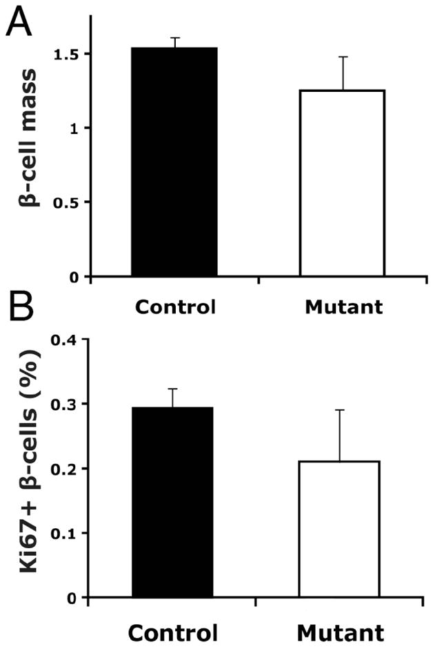
β-cell mass and proliferation in C57Bl/6J Foxm1 mutant and control female mice on HFD. A) β-cell mass was not significantly reduced in Foxm1 mutants at twelve weeks of age (p=0.11). B) The percentage of β-cells positive for the proliferation marker Ki67 was not significantly different between Foxm1 controls and mutants at twelve weeks of age (n=3).