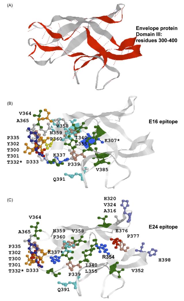
Fig. 4.
Structural comparison of the E16 epitope and predicted E24 epitope. (A) 3D model of domain III of envelope protein. β-strands connecting E16 epitope amino acid contact residues are shown in red. (B) 3D model of epitope E16. (C) 3D model of epitope E24. Colours—red: acidic amino acids, green: hydrophobic amino acids, blue: basic amino acids, orange: serine and threonine. (For interpretation of the references to colour in this figure legend, the reader is referred to the web version of the article.)

