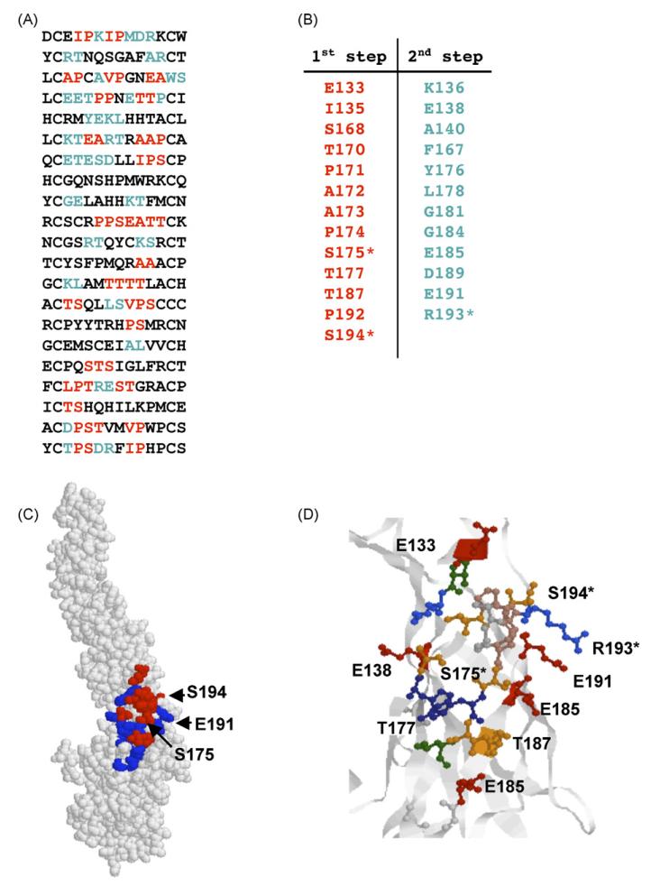
Fig. 5.
Prediction of the E121 epitope. (A) Peptide sequences found by phage library screening. Amino acids predicted at the first stage are shown in red and those predicted at the second step are shown in blue. (B) Amino acids predicted by the algorithm (red) and added at the second step (blue). Amino acids found by mutational analysis are indicated by asterisks. (C) Epitope predicted by our affinity-selected mimotopes. Amino acids predicted at the first stage are shown in red and those predicted at the second step are shown in blue. (D) Folding of predicted E121 epitope. Colours—red: acidic amino acids, green: hydrophobic amino acids, blue: basic amino acids, orange: serine and threonine. (For interpretation of the references to colour in this figure legend, the reader is referred to the web version of the article.)

