Fig. 1.
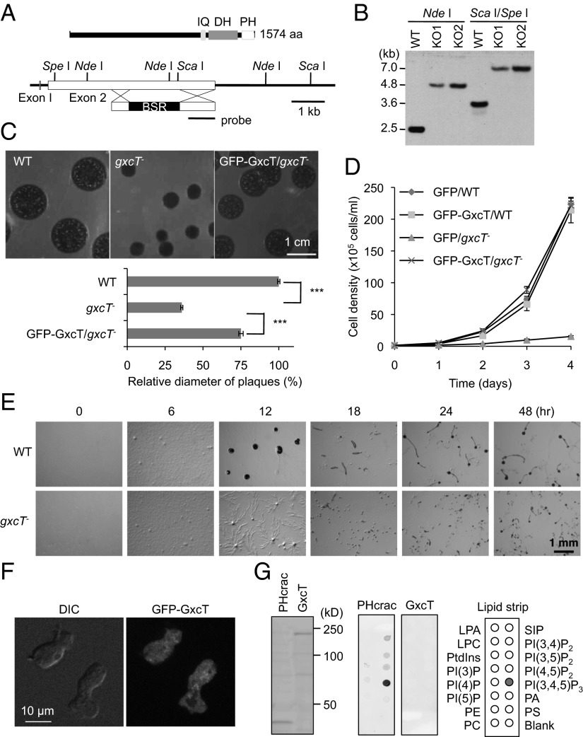
GxcT is necessary for normal cell growth and development. (A) GxcT contains calmodulin-binding (IQ), RhoGEF (DH), and PH domains. The majority of the gxcT gene was replaced by a blasticidin resistance cassette (BSR) flanked by DNA sequences homologous to gxcT. (B) Genomic DNA was digested with the indicated restriction enzymes and analyzed by Southern blotting with a DNA fragment corresponding to the region designated as the probe in A. After digestion with NdeI, WT cells showed a 2.5-kb band as expected, whereas gxcT− cells showed a 4.8-kb band. After digestion with ScaI and SpeI, WT and gxcT− cells showed the expected 3.5- and 7-kb bands, respectively. (C) gxcT− cells formed smaller plaques when grown clonally on bacterial lawns. Expressing a plasmid carrying GFP-GxcT in gxcT− cells nearly restored the WT phenotype (GFP-GxcT/gxcT−). Relative diameter of plaques was quantified (n = 22). (D) WT and gxcT− cells expressing the indicated proteins were cultured in HL5 medium and counted daily with a hemocytometer. Values represent the mean ± SEM (n ≧ 3). (E) WT and gxcT− cells were plated on nonnutrient DB agar and examined over time for development. (F) gxcT− cells expressing GFP-GxcT were observed by fluorescence microscopy. (G) Whole cell lysates were prepared from Dictyostelium cells expressing PHcrac-GFP or GFP-GxcT and immunoblotted using anti-GFP antibodies (Left). Similar whole cell lysates were used for lipid dot blot assays, which showed that GFP-GxcT did not bind any of the indicated lipids (Center and Right).

