Fig. 3.
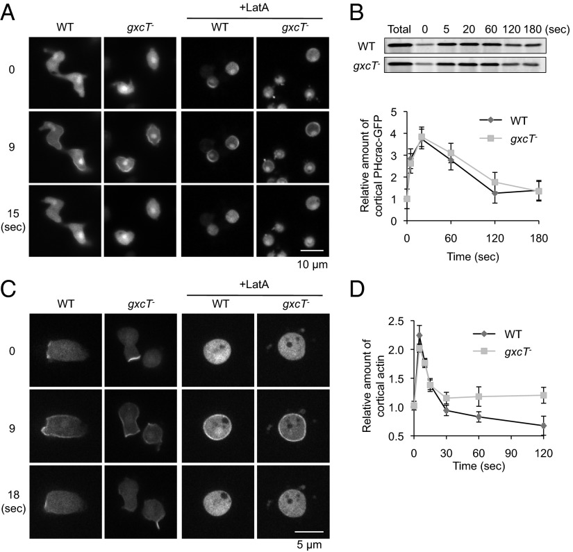
GxcT is dispensible for PIP3 production and Ras activation in response to global chemoattractant stimulation. (A) WT and gxcT− cells expressing the PIP3 biosensor PHcrac-GFP were uniformly stimulated with 1 µM cAMP in the presence or absence of 5 µM Latrunculin A (LatA). Cells were observed by fluorescence microscopy at the indicated time points, relative to the addition of cAMP. (B) At the indicated time points after uniform cAMP stimulation, WT and gxcT− cells expressing PHcrac-GFP were collected and filter lysed. The membrane fraction was isolated and immunoblotted using anti-GFP antibodies (Upper). The total amount of PHcrac-GFP in whole cell lysates is also shown. Band intensity was quantified by densitometry and plotted over time (Lower). Values represent the mean ± SEM (n = 3). (C) Cells expressing a biosensor, RBD-GFP, for Ras activation were uniformly simulated with cAMP in the presence or absence of Latrunculin A (LatA) and observed by fluorescence microscopy as in A. Images are shown for the indicated time points after cAMP addition. (D) At the indicated time points after cAMP stimulation, WT and gxcT− cells were lysed, and the cortical actin was isolated and quantified as described in Experimental Procedures. Values represent the mean ± SEM (n = 3). There are no significant differences between WT and gxcT− cells.

