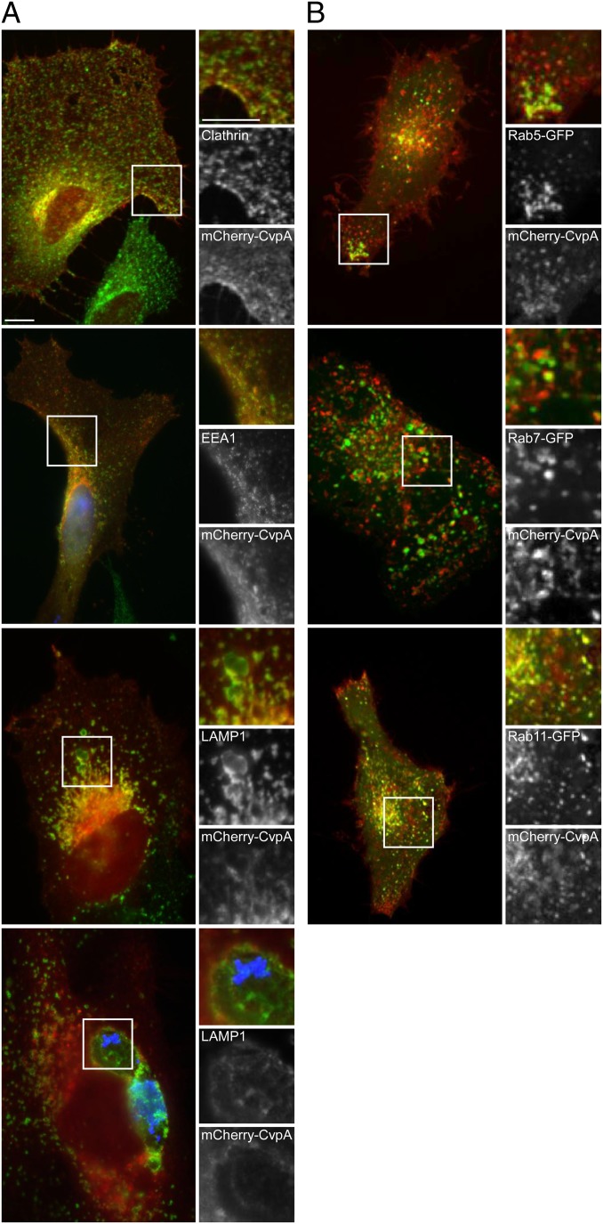
Fig. 3.
Ectopically expressed mCherry-CvpA localizes to endocytic vesicles and traffics to pericentrosomal REs. (A) Representative micrographs of fixed HeLa cells expressing mCherry-CvpA (red) and immunostained for the vesicle proteins clathrin, EEA1, and LAMP1 (green). (Bottom) A cell infected with C. burnetii where bacteria and LAMP1 are immunostained blue and green, respectively. (B) Micrographs of live cells coexpressing mCherry-CvpA (red) and the GFP-tagged Rab GTPases Rab5, Rab7, or Rab11 (green). (Scale bar, 10 μm.)

