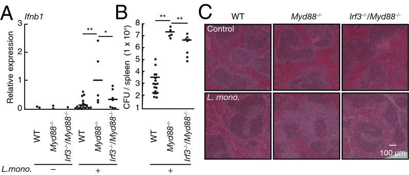
Fig. 4.
Biological significance of TLR-induced suppression of IRF3. (A) Quantitative RT-PCR analysis of IFNb1 mRNA in the spleen from WT (control, n = 2; infected, n = 17), Myd88−/− (control, n = 2; infected, n = 5), and IRF3–MyD88 doubly deficient (Irf3−/−Myd88−/−; control, n = 1; infected, n = 7) mice infected with L. monocytogenes for 2.5 d. **P < 0.01; *P < 0.05. (B) Colony formation assay of L. monocytogenes titers in the spleen of WT (n = 16), Myd88−/− (n = 7), and Irf3−/−Myd88−/− (n = 5) mice infected as in A. Each symbol represents an individual mouse; small horizontal lines indicate the mean. **P < 0.01. (C) Histological analysis of the spleens from WT, Myd88−/−, and Irf3−/−Myd88−/− mice infected as in A and assessed by microscopy of sections stained with H&E. (Magnification: 40×.)

