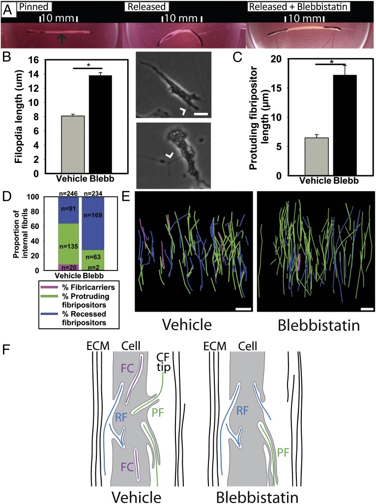
Fig. 6.
NMII is required for fibricarrier formation and normal fibripositor membrane dynamics. (A) Addition of blebbistatin to tendon-like constructs prevented cell-induced shortening when the constructs were unpinned. Arrow (black) shows the tendon-like construct. (B) Protrusive membrane dynamics (such as filopodia formation) are disrupted by blebbistatin. (Upper Right) Nonblebbistatin DMSO control. (Lower Right) Cell in the presence of blebbistatin. Arrowhead shows filopodia. *P ≤ 0.05. (C) Analysis of 3D EM data showed that the membrane protrusion of protrusive fibripositors is longer when NMII is inhibited in embryonic tendon. Bars show the length of the membrane protrusion (*P ≤ 0.05). (D) The proportion of intracellular fibrils in protrusive fibripositors is decreased when NMII is inhibited with blebbistatin for 4 h. Blebbistatin also reduced the proportion of internal fibrils found in fibricarriers. (E) Three-dimensional reconstructions of tendon treated with blebbistatin demonstrate increased numbers of recessed fibripositors (blue) and reduced the numbers of protruding fibripositors (green) with fewer fibricarriers (purple). (F) Schematic representation of the effect of NMII inhibition, showing longer fibripositors and fewer fibricarriers. *P ≤ 0.05. (Scale bars: 5 μm.)

