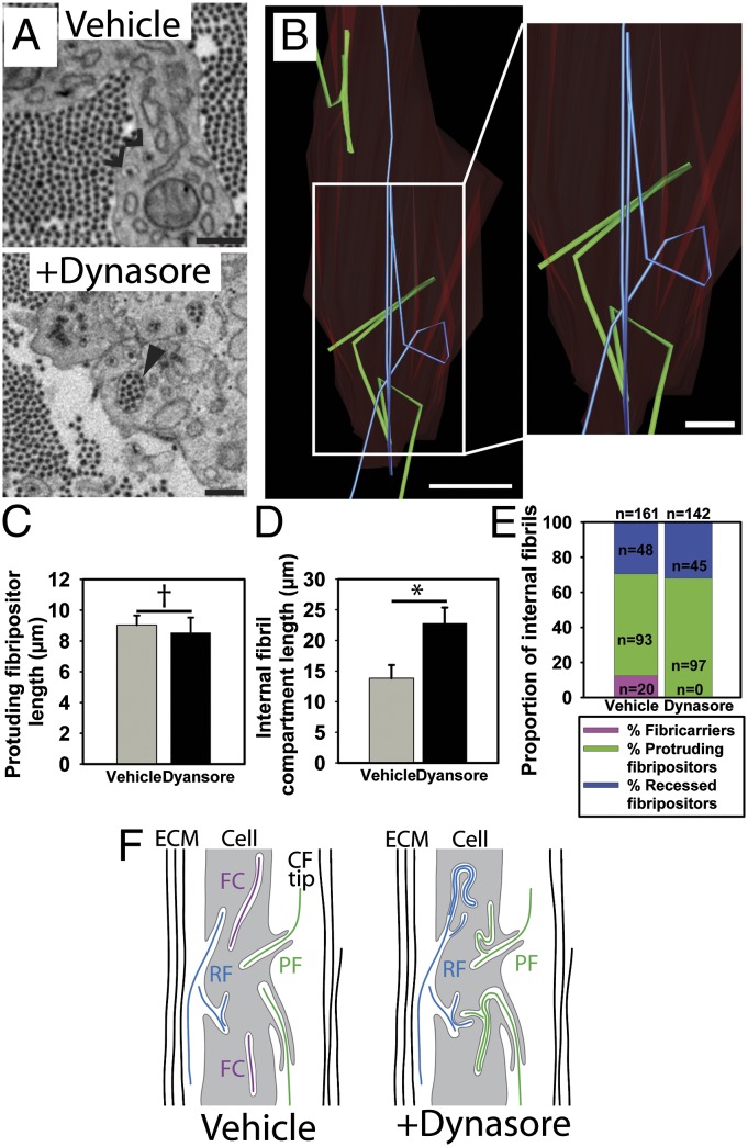
Fig. 7.
Inhibition of dynamin reduces the number of fibricarriers. (A) SBF-SEM images of embryonic chick tendon treated with dynasore demonstrated the appearance of intracellular fibril compartments containing increased numbers of fibrils (marked with a closed arrowhead). Open arrowheads mark recesses containing single fibrils seen in tendon not treated with dynasore. (B) Investigation of this finding in 3D demonstrated that the increased numbers of fibrils seen in 2D cross-sections were the result of internal looping of fibril compartments that were doubled back numerous times. Two different fibrils are shown [in a protruding fibripositor (green) and recessed fibripositor (blue)] illustrating this looping. The cell membrane is colored red. (C) There was no difference in the length of the membrane protrusion of protrusive fibripositors. †P ≥ 0.05. (D) Lengths of internal fibril-containing compartments (recessed fibripositors and the internal part of protruding fibripositors) were significantly increased in dynasore-treated tendon cells. *P ≤ 0.05. (E) No fibricarriers were identified in the 142 fibril compartments studied in 3D in dynasore-treated tissue. In contrast, 20 fibricarriers were identified in 161 fibril compartments studied in untreated tissue. (F) Schematic showing looped internal fibril recesses and reduced number of fibricarriers after dynasore treatment. (Scale bars: A, 500 nm; B Left, 5 μm; B Right, 2 μm.)

