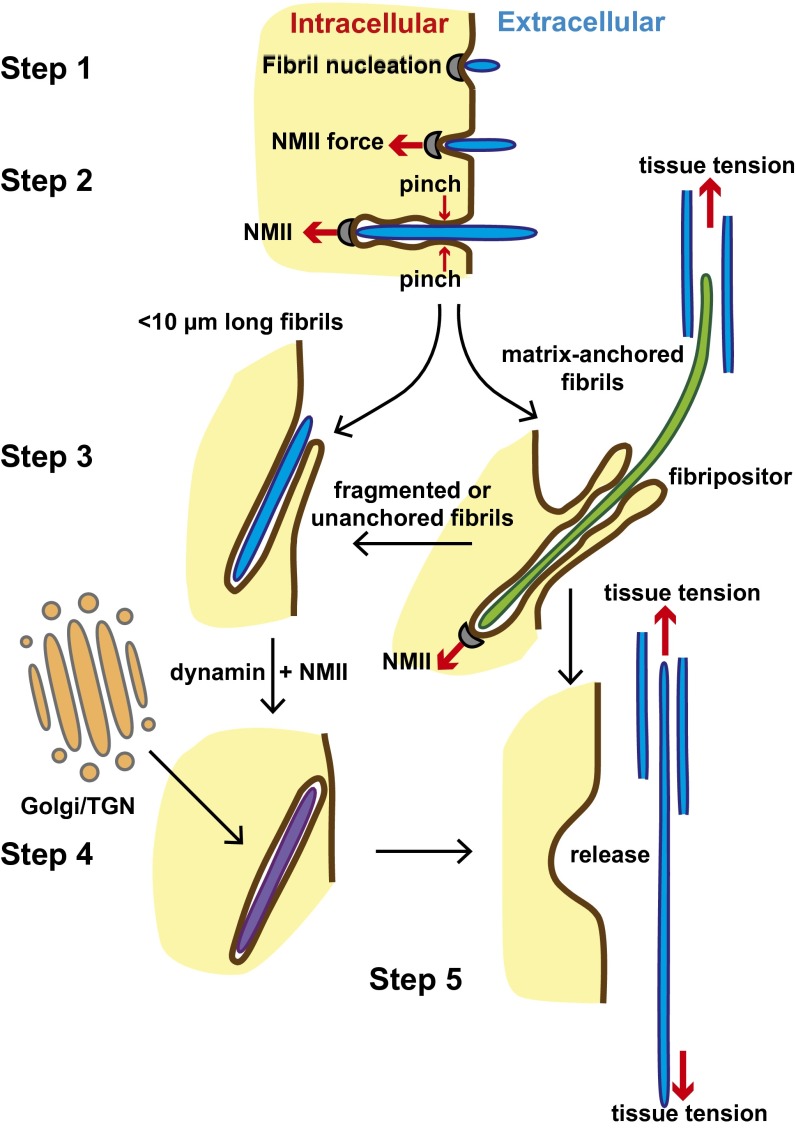
Fig. 9.
Model of collagen fibril nucleation and transport. Schematic represents the processes of collagen fibril nucleation and movement between the experimentally-observed compartments. The transportation process is driven primarily by NMII-generated forces. The cell membrane is brown, the cytosol is yellow, and the extracellular matrix is white. Fibrils in protruding fibripositors are green, those in recessed fibripositors are blue, and those in fibricarriers are purple. Collagen fibrils are formed at the cell surface by nucleation of collagen (step 1). Newly formed fibrils are pulled into a membrane recess by NMII (step 2). Fibrils continue to grow by molecular accretion, and tension is continually exerted on these fibrils by NMII. Long fibrils contained in protruding fibripositors are positioned in the matrix, whereas short fibrils remain in recessed fibripositors (step 3). Fragmented or unanchored fibrils may be totally internalized in a process requiring NMII and dynamin, forming fibricarriers. Fibrils inside fibricarriers may grow by secretion of collagen from the Golgi/TGN apparatus (step 4). Fibricarriers secrete their fibril cargo back into the ECM (step 5). We propose that the tension exerted on matrix-anchored fibrils in fibripositors by NMII-generated cell force is important in generating an aligned tissue that is tensioned.

