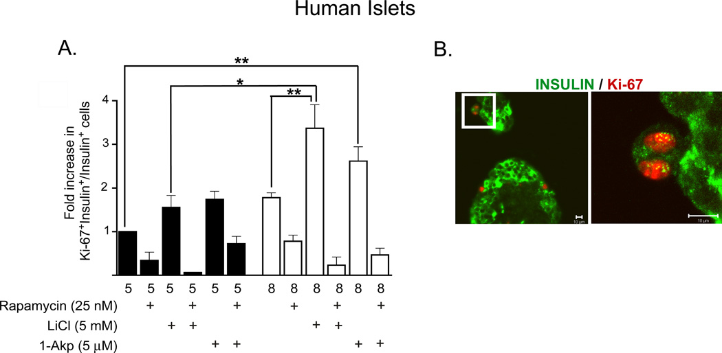
Figure 8. Lithium and 1-Akp in combination with glucose stimulates β-cell proliferation in human islets.
(A) Fold increase (with respect to 5 mM glucose) in the ratio of proliferating β-cells to total number of insulin+ β-cells as determined by immunohistochemical analysis. Data presented are the mean ± SE of three independent experiments. For each group per experiments, ~5,000–6,000 β-cells were counted. Glucose (8 mM) + LiCl significantly increased the number of Ki-67+/insulin+ cells compared to that of 8 mM glucose alone or 5 mM glucose + LiCl. A 4-fold increase in proliferating β-cells was noted in human islets treated with 8 mM glucose + LiCl (0.71%+ 0.3) in contrast to 5 mM glucose + LiCl (0.168% + 0.08) or 8 mM glucose alone (0.34& ± 0.2). Rapamycin treatment resulted in significantly fewer Ki-67+/insulin+ cells in all treatment conditions. *P < 0.05, **P < 0.01, and ***P < 0.001 denote significant differences between the bracketed conditions. (B) Representative immunofluorescent images of human β-cells showing β-catenin cellular localization. a: insulin positive (green cytoplasm), nuclear β-catenin positive (purple nucleus). b: insulin positive (green cytoplasm), nuclear β-catenin negative (blue nucleus). c: insulin negative, nuclear β-catenin positive (purple nucleus; red nuclear ring). d: insulin negative, nuclear β-catenin negative (blue nucleus). Reprinted with permission from ref. [57].

