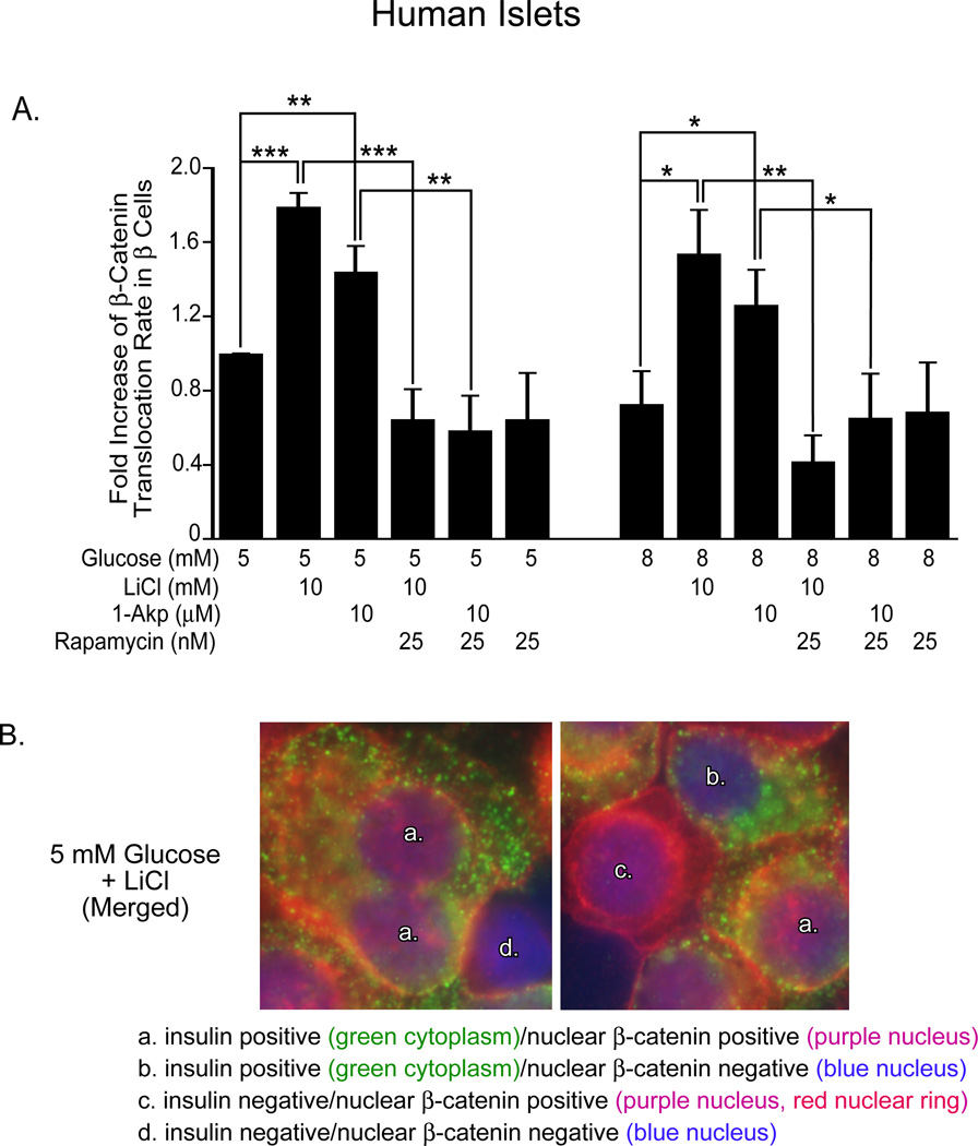
Figure 9. Translocation of nuclear β-catenin in human islets.
(A) Fold increase in β-catenin nuclear positive/insulin positive cells (with respect to 5 mM glucose) as determined by immunohistochemical analysis. Data are the means ± S.E. of n = 3 experiments. For each group per experiment, ~5,000–6,000 β-cells were counted. *p<0.05, **p<0.01, and ***p<0.001 denote significant differences between the bracketed conditions. (B) Representative immunofluorescent images of dispersed human islets to demonstrate co-localization of cytoplasmic insulin and nuclear β-catenin used for quantitation in Figure 8A. Reprinted with permission from ref. [57].

