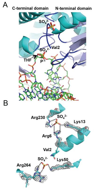
Figure 2.
The sulfate ions in the unliganded MvNei2 structure likely mimic DNA backbone phosphates. (A) Superposition of unliganded MvNei2 with the DNA from the MvNei1-THF structure (PDB ID 3A46 [27], performed with SSM using all atoms in COOT [41]), indicates that one of the two sulfates found in the MvNei2 structure lies in the same position as a phosphate in the DNA. MvNei2 is colored and positioned as in Fig. 1A. The sulfate ions are colored by atom type, MvNei1-DNA carbon atoms are shown in green and the bases and backbone phosphates are colored by atom type. (B) Coordination of the sulfate ions by basic lysine and arginine residues at the surface of the MvNei2 enzyme. The enzyme is colored in cyan with the side chains and the sulfates colored by atom type. A composite omit map (PHENIX [43]; grey) contoured at 1σ is overlaid on the residues coordinating the sulfate ions.

