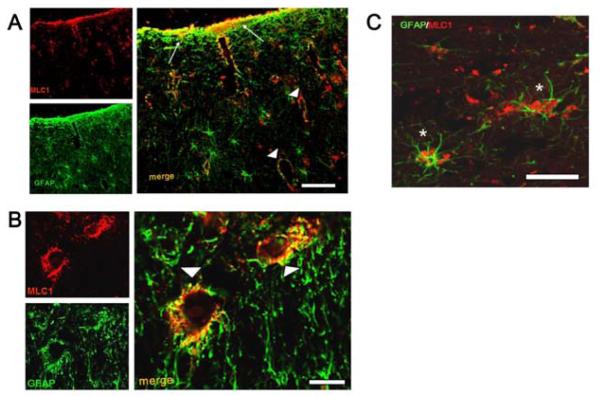
Figure 8. Co-immunostaining of normal human brain with anti-MLC1 (red) and anti-GFAP (green) antibodies.
MLC1 is expressed in astrocyte end-feet contacting pial membrane (A, arrows) and blood vessels (arrowheads in A and in B) as indicated by the yellow signal in merged images. Scattered astrocytes in the parenchyma show MLC1 staining in vesicular structures in the cell body (C, asterisks). Bars A=50 μm; B,C=20 μm. (Modified from Ambrosini et al., Mol. Cell. Neurosc., [177]).

