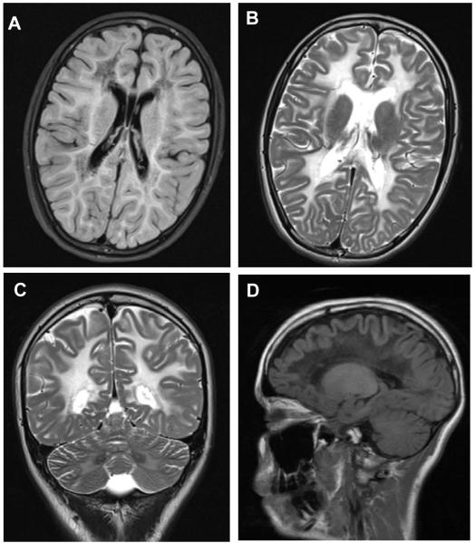
Figure 9. MRI of a 14 year old girl with 2 heterocompound mutations in eIF2Bε.
(A) Notice the diffuse hypointensity of the abnormal signal in FLAIR weighted image sparing the U fibers indicating abnormal vacuolation of the white matter; (B) the same areas appear markedly hyperintense in the T2 weighted images and also involve the internal capsule; (C) the abnormal hyperintense T2 weighted signal also involves the white matter of the cerebellum; (D) the increased vacuolation is also clear in the parasaggital FLAIR weighted image showing a marked hypointense signal of the brain white matter.

