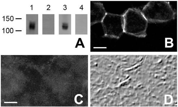
Figure 1.
A: Immunoblots, probed with the NDCBE-1C antibody: Lanes 1 and 2, brain lysate; Lanes 3 and 4, lysate from HEK cells expressing NDCBE-A. Preadsorption of the antibody with the corresponding peptide (lanes 2 and 4) blocked the signal. B: Immunofluorescence labeling for NDCBE-1r in HEK 293 cells transfected with NDCBE-A. C: Immunofluorescence labeling for NDCBE-1r in HEK 293 cells transfected with NDCBE-A in the presence of blocking peptide. D: Field in C, visualized by differential interference contrast imaging. Scale bar = 5 μm in B (also applies to D) and C.

