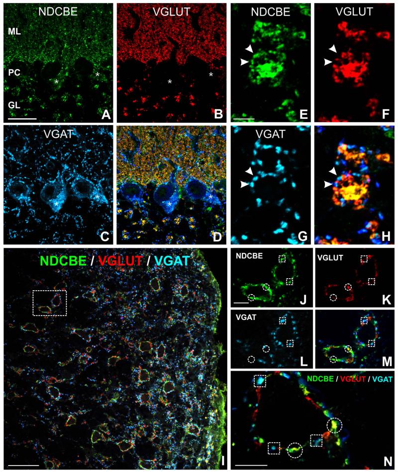
Figure 5.
Co-localization of NDCBE (green) with VGLUT1 (red) and VGAT (blue) in rat hindbrain. A-D: In cerebellar cortex, VGLUT puncta exhibit considerable co-localization with NDCBE; note co-localization between VGAT and NDCBE in the Purkinje cell layer. E-H: Synaptic glomeruli in granule cell layer exhibit a characteristic pattern of co-localization: NDCBE is associated both with the large central VGLUT-positive terminal and with the surrounding VGAT-positive inhibitory terminals. I: Cochlear nucleus. J-N: Co-localization pattern in more detail (J-M are from boxed region in I). Many NDCBE puncta co-localize with VGLUT (circles), whereas others co-localize with VGAT (boxes).Scale bar = 25 μm in A (applies to A-D); 5 μm in E (applies to e-H); 50 μm in I; 10 μm in J (applies to J-M) and N.

