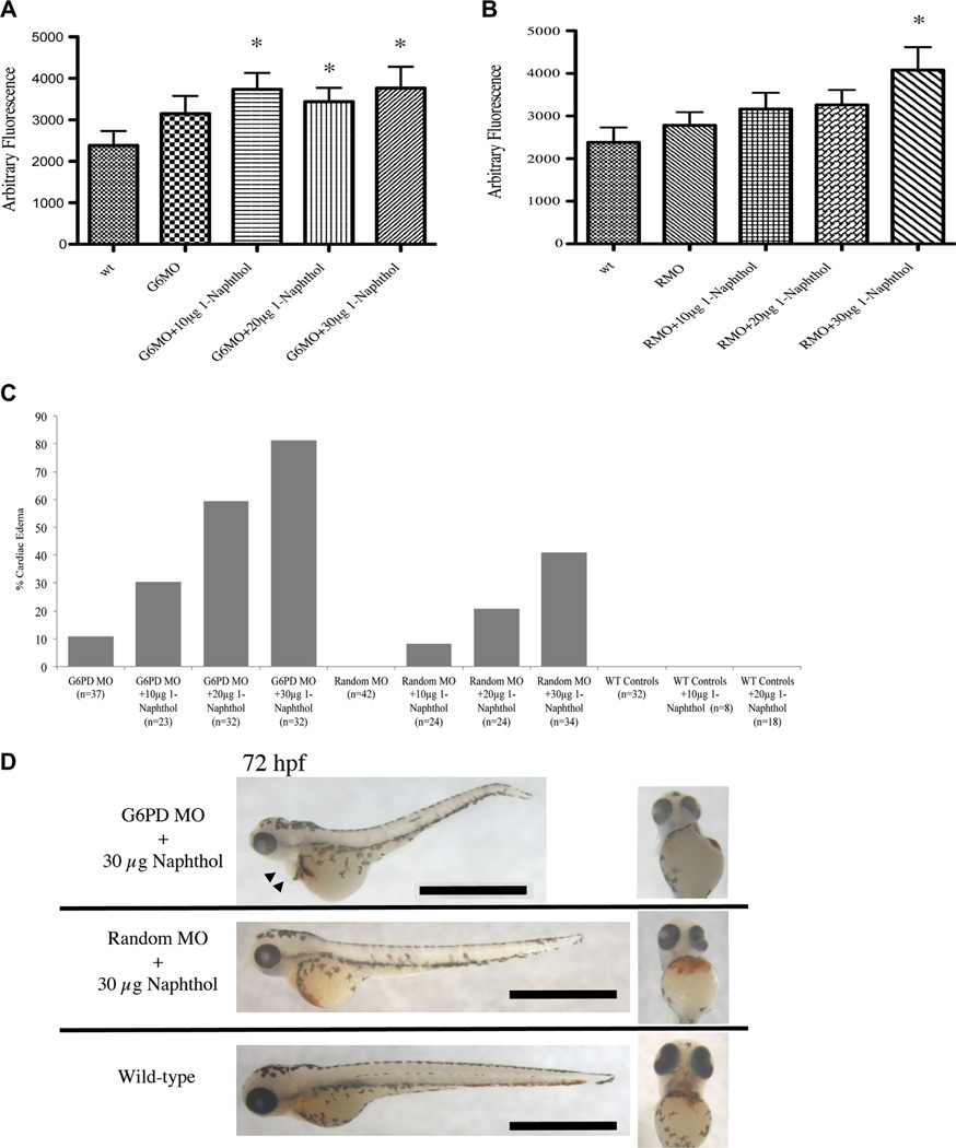
Figure 5.
1-Naphthol induces oxidative stress and edema in g6pd morphants. (A, B)Wild type embryos were injected with 1.2 pmol g6pd MOs or random MOs followed by exposure to increasing amounts of 1-naphthol per 5mL of embryo water from24 to 72 hpf. Embryos were placed in a 96-well plate, and CM-H2DCFDA was added to 500 ng/mL. Fluorescence activity was read using 485/20 nm excitation and 528/20 nm emission on a Biotek plate reader. Shown are the mean and SEM (n = 21–29 embryos per group). *p = 0.01, Student t test compared with the wild type group. (C) Numbers of embryos with pericardial edema were tallied after exposure to 10–30 µg 1-naphthol. Data were subjected to χ2 analysis to generate p < 0.0001. (D) MO-injected, 1-naphthol–treated zebrafish were stained with o-dianisidine to show hemoglobin-producing cells at 72 hpf. Black triangles indicate the extent of the pericardial edema. The scale bars represent 1000 µm.

