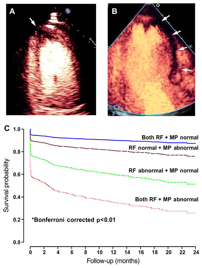
Figure 1.

Bedside diagnosis of acute coronary syndrome. The top images are examples of acute myocardial infarction from two patients detected by bedside myocardial contrast echocardiography perfusion imaging where regions of hypoperfusion (arrows) are depicted by lack of myocardial opacification. The images illustrate (A) a small risk area in the apical four chamber view, and (B) a large risk area in the apical three chamber view. (C) Adjusted event-free survival (myocardial infarction, death, and heart failure) in 1017 patients presenting to the emergency department with chest pain undergoing myocardial contrast echocardiography.2 Data are stratified according to whether regional function (RF) and myocardial perfusion (MP) were normal or abnormal and comparisons made with Cox proportional hazards model.
