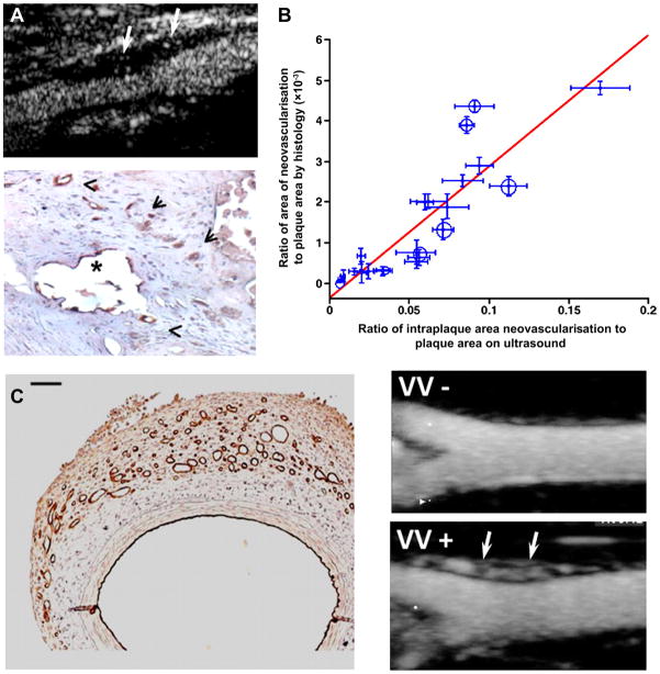
Figure 3.
Contrast enhance ultrasound (CEU) imaging of vasa vasorum (VV) and plaque neovascularisation. (A) Examples of plaque neovascularisation detected by CEU in a patient with severe carotid stenosis (top) and also by histology with dual CD31/CD34 immunostaining after endarterectomy (bottom).6 (B) Relation between plaque neovascularisation by histology of carotid endarterectomy specimens and by CEU before surgery.w17 Black circles are from symptomatic patients and scales for x and y axis data are different. (C) Examples of VV proliferation produced by a model of vascular haemorrhage detected by CD31 immunohistology (left) and CEU with maximal intensity projection imaging of a rabbit femoral artery in long axis without (VV−) and with (VV+) VV proliferation (arrows) produced by haemorrhage.7

