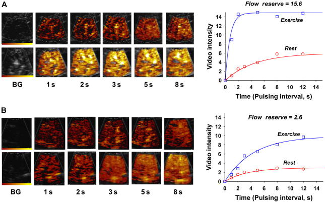
Figure 4.
Contrast enhanced ultrasound (CEU) images and corresponding time-intensity data obtained from the plantar flexor calf muscles at rest and during plantar flexion exercise in (A) a control subject and (B) a patient with moderate peripheral arterial disease in whom there is a pronounced reduction in hyperaemic flow and flow reserve.8 Images and data were obtained after a destructive pulse sequence. BG, background.

