
Figure 2.
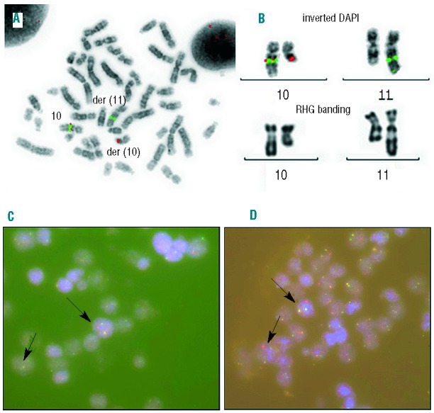
Molecular and conventional cytogenetics of bone marrow in the acute myelomonocytic leukemia stage (A and B) and paraffin embedded pleural fluid pellet in the T-cell lymphoblastic lymphoma (C and D). (A) Fluorescent in situ hybridization (FISH) analysis using BACs RP11-9E13 (spectrum orange) and RP11-314J18 (spectrum green, BlueGnome®) flanking TET1gene that are separated in the t(10;11)(q22;q23) translocation. (B) Magnification of the chromosomes 10 and 11 in inverted DAPI and XL MLL break apart probe (Metasystems®) FISH and RHG banding, demonstrating the rearrangement of MLL. (C) FISH study using an XL MLL break apart probe in pleural effusion. A split of the MLL probe had been observed within the malignant cell population (arrows). (D) Interphasic FISH study of TET1 using BACs probes RP11-9E13 (spectrum orange) and RP11-314J18 (spectrum green). A separation of the signals for one allele had been observed within the malignant cell population.