Figure 1.
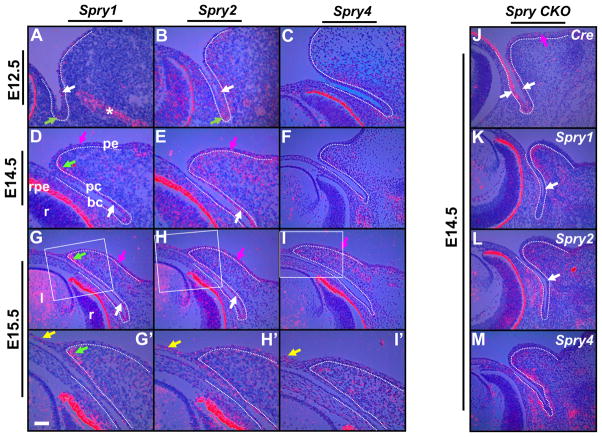
Spry 1, Spry2 and Spry4 expression in wildtype and Spry CKO mutant eyelids. 35S-labeled Spry1 (A, D, G, G′, K), Spry2 (B, E, H, H′, L), Spry4 (C, F, I, I′, M) and Cre (J) riboprobes were hybridized to sections of E12.5 (A, B, C), E14.5 (D, E, F) and E15.5 (G–I′) wildtype and E14.5 (J–M) Spry CKO mutant embryos. The dotted lines delineate the boundary between the epithelial and mesenchymal cells. G′–I′ are higher magnifications of boxed regions in G–I. Conjunctival epithelial cells expressed Spry1 (A, D, G, white arrows) and Spry2 (B, E, H, white arrows), the palpebral epidermal cells expressed Spry1 (D, G, magenta arrows), Spry2 (E, H, magenta arrows) and Spry4 (I, magenta arrow) and the eyelid mesenchymal cells showed localized expression of Spry1 (D, G, G′, green arrows) but broader expression of Spry2 (E, H, H′) and Spry4 (F, I, I′). Peridermal cells expressed all three Sprys (G′–I′, yellow arrows). In the Spry CKO eyelids, the conjunctival epithelial cells expressed Cre recombinase (J, white arrows) and showed near complete loss of Spry1 (K, white arrow) and Spry2 (L, white arrow) expression. The eyelid mesenchymal cells showed increased expression of Spry1 (compare K to D) and Spry 4 (compare M to F) and the bulbar mesenchymal cells showed increased expression of Spry2 (compare L to E) and Spry4 (compare M and F). Asterisk in panel A marks Spry1 expression in the nasolacrimal duct. Staining in the retinal pigmented epithelium (D, rpe) is an artifact of dark-field illumination. Abbreviations; bc, bulbar conjunctiva; l, lens; pc, palpebral conjunctiva; pe, palpebral epidermis; r, retina. Scale bar in (G′): (A, B, C, G′–I′) 50 μm; (D–I, J–M) 100 μm.

