Figure 5.
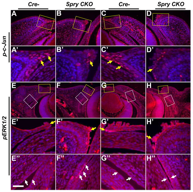
ERK and c-Jun phosphorylation in Spry CKO eyelids. Alterations in ERK and c-Jun phosphorylation in E14.5 Cre- and Spry CKO eyelids were analyzed immunohistochemistry. Upper eyelids are shown in panels A–B′, E–F″ and lower eyelids are shown in panels C–D′, G–H″. A′–H′ are higher magnifications of anterior margins (yellow squares) in panels A–H. E″–H″ are higher magnifications of conjunctiva (white squares) in E–H. c-Jun phosphorylation is reduced in the peridermal cells at the leading edge of Spry CKO upper (B′, arrow) and lower (D′, arrow) eyelids. ERK phosphorylation in the Spry CKO eyelids (F′, H′) remained unaltered in the peridermal cells. In contrast, conjunctival epithelial cells of Spry CKO upper (F″, arrows) and lower (H″, arrows) lids showed increased ERK phosphorylation. Scale bar in (E″): (A–D) 120 μm; (A′–D′) 40 μm; (E–H) 240 μm; (E′–H″) 40 μm.

