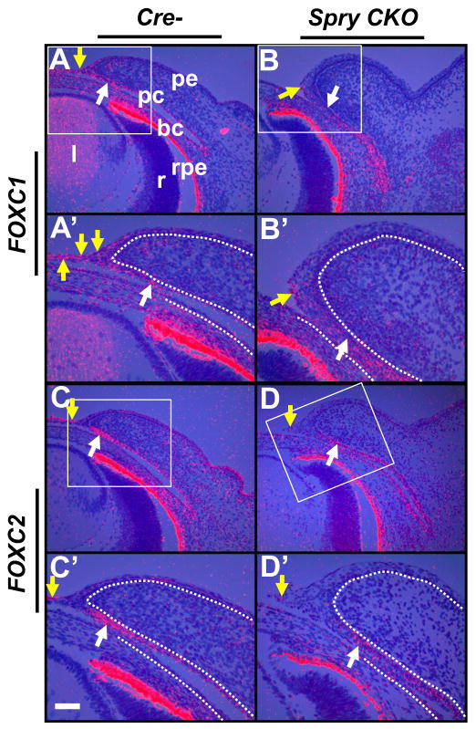
Figure 7.
Foxc1 and Foxc2 expression in Spry CKO eyelids. 35S-labeled Foxc1 (A–B′) and Foxc2 (C–D′) riboprobes were hybridized to sections of E14.5 Cre- and Spry CKO mutant embryos. The dotted lines delineate the boundary between the epithelial and mesenchymal cells. A′–D′ are higher magnifications of boxed regions in A–D. Modest reduction in Foxc1 (B, B′) and a strong reduction in Foxc2 (D, D′) was seen at the anterior margins of Spry CKO eyelids when compared to Cre- eyelids (B, B′, white arrows). Spry CKO peridermal cells also showed reduced expression of Foxc1 (B′, yellow arrow) and Foxc2 (D, D′, yellow arrow) compared to Cre- eyelids (A, A′, C, C′, yellow arrows). Staining in the retinal pigmented epithelium (rpe) is an artifact of dark-field illumination. Abbreviations; bc, bulbar conjunctiva; l, lens; pc, palpebral conjunctiva; pe, palpebral epidermis; r, retina. Scale bar in (C′): (A–D) 100 μm; (A′–D′) 50 μm.

