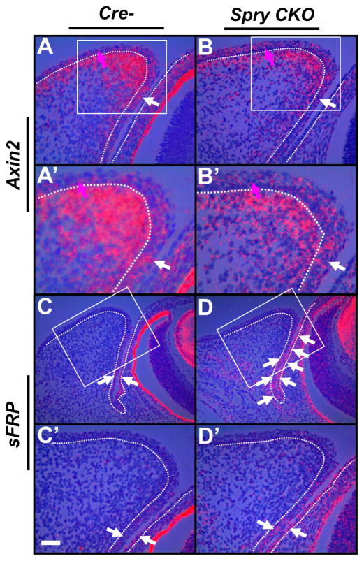
Figure 8.
Decreased Wnt signaling in Spry CKO eyelids. 35S-labeled Axin2 (A–B′) and sFRP1 (C–D′) riboprobes were hybridized to sections of E14.5 Cre- and Spry CKO mutant embryos. The dotted lines delineate the boundary between the epithelial and mesenchymal cells. A′–D′ are higher magnifications of boxed regions in A–D. Axin2 (B, B′) expression was modestly reduced in the Spry CKO conjunctival epithelial cells (B, white arrow) and in eyelid mesenchymal cells (A, B) but was not in the palpebral peridermal cells (B, magenta arrows). sFRP1 was increased in the Spry CKO conjunctival epithelial cells (D, D′, white arrows). Staining in the retinal pigmented epithelium (rpe) is an artifact of dark-field illumination. Abbreviations; bc, bulbar conjunctiva; l, lens; pc, palpebral conjunctiva; pe, palpebral epidermis; r, retina. Scale bar in (C′): (A, B, A′, B′, C′, D′) 50 μm; (C, D) 100 μm.

