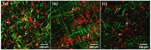
Figure 3.

Composite z-stack confocal images showing spreading and arrangement of phalloidin labeled cells highlighting F-actin (red) on day 0 (a) Pμ (b) PμPn and (c) PμFn scaffolds (green). Arrows indicate examples of individual cell morphologies and arrangements.
