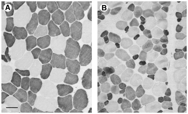
FIGURE 4.

Mean fiber area differences compared between a deep bite and normal bite malocclusion subject. Sections of masseter muscle immunostained with antibody to fast (type II) myosin (Sigma clone my32), photographed at the same magnification (scale bar = 50 μm). A, Deep bite patient, B, normal bite patient. Strongly stained fibers are type II; pale stained are type I. Note the difference in type II fiber areas in A compared with B, and the large population of intermediate stained fibers in B, many of which were type I/II hybrid fibers.
Sciote et al. Human Masseter Muscle Fiber Type Properties. J Oral Maxillofac Surg 2012.
