
Figure 11.
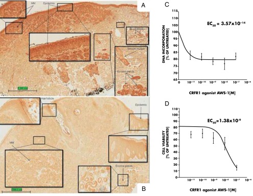
Expression of CRF and CRFR-1 in the normal skin, melanoma cells, and effects of CRF1 agonist on proliferation of melanoma cells. A, CRF1 is expressed in normal structures of human skin such as epidermis, blood vessels, eccrine glands, and smooth muscle as well as in malignant melanoma cells (MM). B, CRF is expressed in normal structures of human skin such as epidermis, eccrine glands, and hair follicle as well as in malignant melanoma cells (MM). The slides in A and B were stained with antibodies as described previously (209). Magnification, ×20; insets, ×200. C, CRFR-1 agonist AWS-1 inhibits proliferation of AbC1 hamster melanoma cells. Cells were incubated with the peptide for 48 hours in the 154 medium (Cascade Biologics, Inc) containing growth factors. The DNA synthesis was measured with titrated thymidine incorporation, and data were analyzed as described previously (117). D, CRFR-1 selective agonist AWS-1 inhibits proliferation of Melan A mouse immortalized melanocytes. Cells were incubated with the peptide for 48 hours in the F10 medium containing fetal calf serum (Invitrogen, Inc). The cell viability was measured with 3-(4,5-dimethylthiazol-2-yl)-2,5-diphenyltetrazolium bromide (MTT) assay, and data were analyzed as described previously (117). The difference between control and treatments was analyzed with one-way ANOVA (P < .005) as described previously (117). Panels A and B were prepared by Dr Diane Kovacic, a dermatopathology fellow at the University of Tennessee Health Science Center.