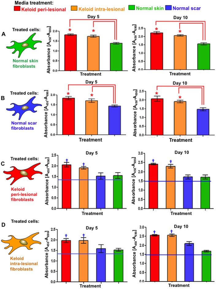
Figure 5. Proliferation after 120(day-5) and 240 hrs (day-10) of conditioned media treatment.
A. Significantly increased (*p<0.03) proliferation and cellular viability was observed in both NF and NS treated with PKF or IKF media versus respective control media after 120 hrs. B. Similar trends were observed after 240 hrs although overall proliferation levels were higher than corresponding treatments at 120 hrs. C. Significantly higher proliferation was observed in PKF and IKF when treated with PKF or IKF media versus NF or NS media after 120 hrs. D. Similar trends were observed for PKF and IKF cells at 240 hrs with overall proliferation higher than corresponding treatments at 120 hrs. NF = normal dermal fibroblasts (n = 4), NS = Normal dermal scar fibroblasts (n = 4), PKF = peri-lesional keloid fibroblasts (n = 5), IKF = intra-lesional keloid fibroblasts (n = 5). Significantly increased (†p<0.02) proliferation and cellular viability was also observed in both PKF and IKF treated with PKF or IKF media versus respective NF and NS when treated with NF and NS media.

