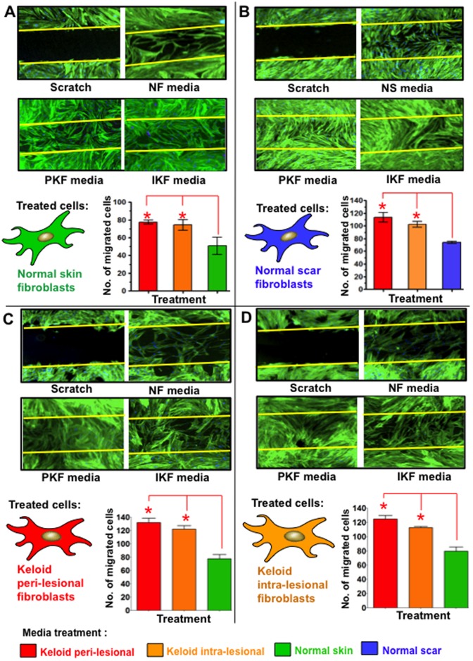
Figure 6. Cell migration in scratch wound invasion assay after 240 hrs of conditioned media treatment.
A. Significantly increased (*p<0.03) cell migration occurred in NF over a 30 hrs period following 240 hrs treatment with PKF or IKF conditioned media versus NF control media. B. Significantly increased migration into a scratch wound was also observed in NS treated with PKF and IKF versus NS control media. C. PKF treated with NF control media formed a mesh-like network of cells in the scratch wound and significantly (p<0.01) increased migration following treatment with PKF and IKF conditioned media as compared to normal skin fibroblasts condition media. D. Significant (p<0.03) increased migration was also observed in IKF cells following treatments with PKF or IKF media versus NF control media. Blue = nuclei, Green = phalloidin stained intermediate filaments. NF = normal dermal fibroblasts (n = 4), NS = Normal dermal scar fibroblasts (n = 4), PKF = peri-lesional keloid fibroblasts (n = 5), IKF = intra-lesional keloid fibroblasts (n = 5).

