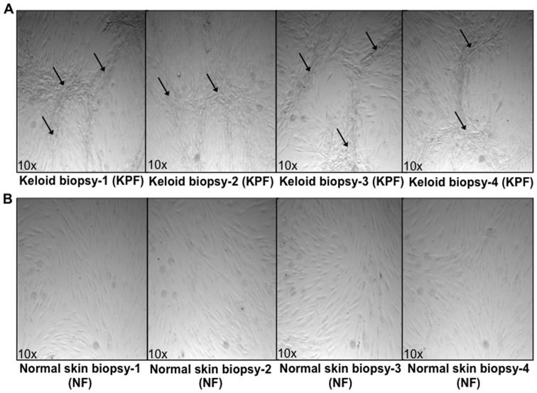
Figure 7. Cellular organisation in post-confluent in vitro primary cultures.
A. Keloid primary fibroblasts derived from the peri-lesional margin of four keloid scar biopsies (PKF) passage ≤3. Arrows indicate whirl-like ridges and nodular aggregates formed when cells were grown to a post-confluent (>100%) state. B. Normal skin primary fibroblasts (NF) grown to a post-confluent (>100%) state did not show ridge structures or aggregates but grew in parallel layers.

