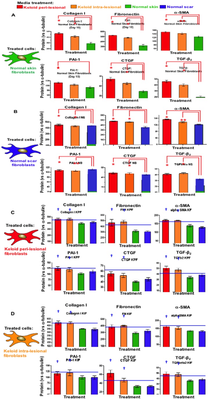
Figure 8. Protein expression after 240 hrs of replenishing conditioned media treatment.
A. Significantly increased (*p<0.04) expression of collagen I, fibronectin, α-SMA, CTGF, PAI-1 and TGFβ-2 was observed in NF treated with PKF or IKF conditioned media versus NF control media after 240 hrs. Representative triplicates are shown below graphs (green) normalised against α-tubulin (red). B. Increased expression was observed for NS treated with PKF or IKF media versus NS control media after 240 hrs. C. PKF treated with PKF or IKF media elicited higher expression of all protein markers in comparison to both NF and NS media at 240 hrs. D. IKF treated with PKF or IKF media also increased expression compared to both NF and NS media at 240 hrs. NF = normal dermal fibroblasts (n = 4), NS = Normal dermal scar fibroblasts (n = 4), PKF = peri-lesional keloid fibroblasts (n = 5), IKF = intra-lesional keloid fibroblasts (n = 5). Significantly increased (†p<0.04) expression of collagen I, fibronectin, α-SMA, CTGF, PAI-1 and TGFβ was also observed in both PKF and IKF treated with PKF or IKF media versus respective NF and NS when treated with NF and NS media.

