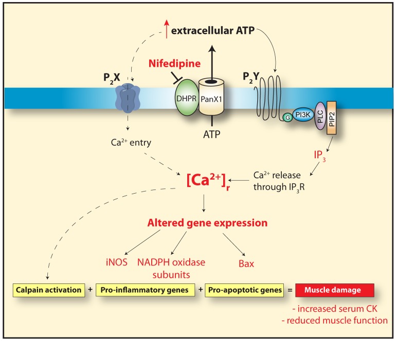
Figure 9. Proposed model for ATP-mediated effects in dystrophic skeletal muscle.
In dystrophic skeletal muscle fibers there is an increase in basal ATP release, through Pannexin1 channels that is modulated by the DHPR [12]. Extracellular ATP increases the [Ca2+]r in dystrophic muscle fibers through the activation of purinergic receptors (P2X, inotropic and P2Y, metabotropic). This leads to the expression of pro-apoptotic and pro-inflammatory genes, increasing the muscle damage observed in dystrophic skeletal muscle cells. Nifedipine treatment reduces the basal ATP release and reduces [Ca2+]r, resulting in less pro-inflammatory and pro-apoptotic gene expression and subsequently reduces muscle damage as indicated by a decrease in blood CK and an increase in muscle function assessed by the inverted grid-hanging test and the force swimming test.

