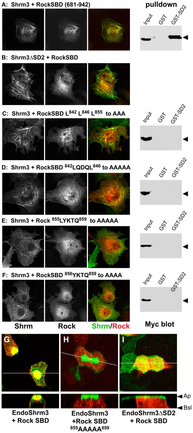
Figure 5. The Rock1 SBD is required for localization with Shrm3.
(A–F) Myc-tagged wild type or SBD variants of hRock1 (681–942) were co-expressed with wildtype Shrm3 or a Shrm3 variant lacking the SD2 in Cos7 cells and stained to detect Shrm3 (green) or the myc tag (red). The right-hand panels in A, C–F depict the results of pulldown assays to detect the interaction of the indicated SBD variant and the Shrm3 SD2. Binding of the Rock SBD variants was tested by using immobilized GST-Shrm3 SD2 and lysates from HEK293 cells expressing the indicated SBD protein, followed by western blotting to detect the myc-tagged SBD proteins. Input = total cell lysate, GST = pulldown using GST bound to beads, GST-SD2 = GST-Shrm3-SD2 bound to beads. Arrowhead denotes the myc-tagged Rock protein. (G–I) T23 MDCK epithelial cells were transfected with expression vectors for EndoShrm3 and Rock1 SBD (G), EndoShrm3 and Rock1-SBD 855LYKTQ859 to 855AAAAA859 (H) or EndoShrm3ΔSD2 and Rock1-SBD (I), grown on transwell filters overnight to form confluent monolayers, and stained to detect EndoShrm3 (green) and Rock-SBD (red). Dashed lines indicate the position of the Z-projections that are shown in the lower panels. Ap, apical surface; Bsl, basal surface.

