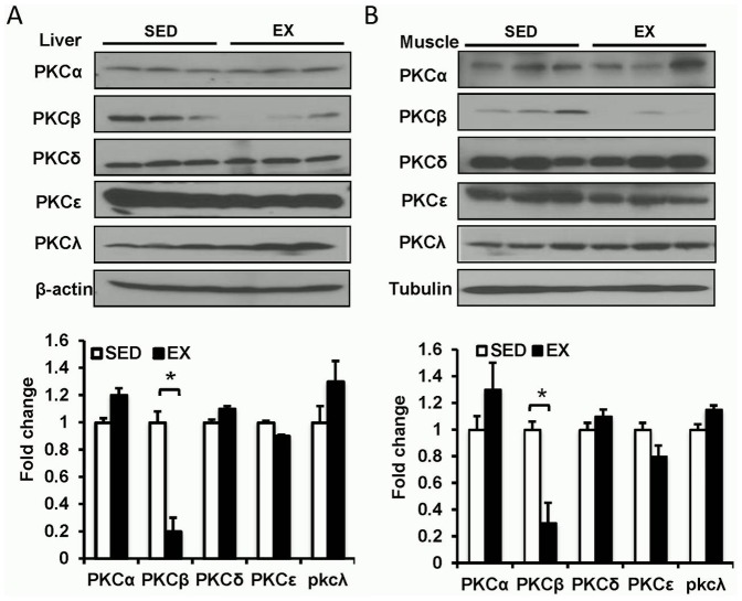
Figure 1. Expression of PKC isoforms in HFD and exercised mice.
A, Expressions of PKC isoforms in liver. Wild-type (WT) mice fed on a high-fat diet (HFD) feeding were either exercised (EX) or sedentary (SED) for 8 weeks. Liver was isolated and used for Western blot detection of PKC isoforms (Top panel, representative images; bottom panel, statistical analysis). *, P<0.05. B, Expressions of PKC isoforms in skeletal muscle. WT mice fed on a HFD were either exercised or sedentary for 8 weeks. Skeletal muscle tissue was isolated and used for Western blot detection of PKC isoforms (Top panel, representative images; bottom panel, statistical analysis). *, P<0.05.

