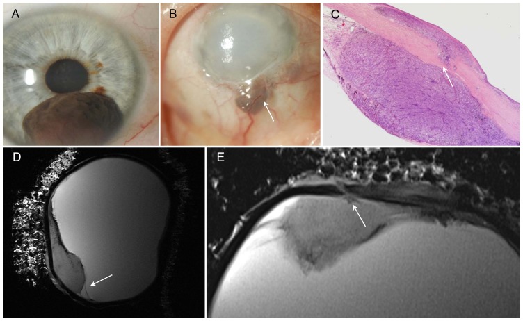
Figure 2. Malignant melanoma of iris and ciliary body with scleral perforation two years after iridocyclectomy.
A – Photograph: malignant melanoma of iris at 6 o'clock before surgery (iridocyclectomy). B – Photograph: recurred tumor with scleral perforation at 6 o'clock, vascularization and corneal edema (arrow). C – H&E stain, 2× magnification: tumor extends through the sclera (arrow). Conjunctiva demonstrates mushroom-like detachment. D – Sagittal T2w image: tumor with hypointens rim (paramagnetic effect of melanin) and retinal detachment (arrow). E – Axial T2w image: delineation of the melanoma from the bulbar wall and good visualization of the perforation (arrow).

