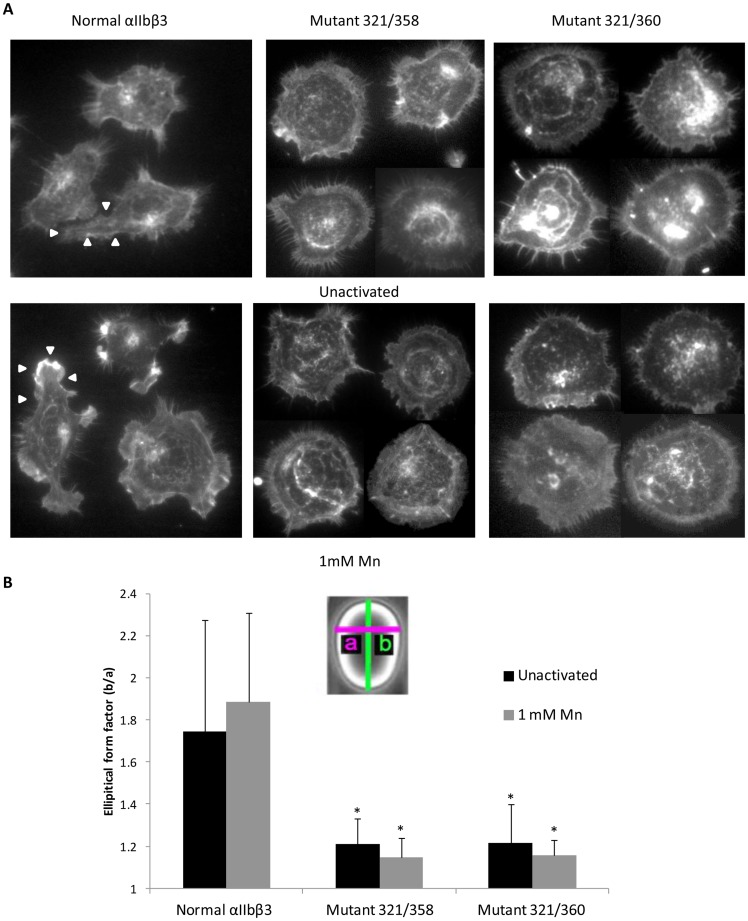
Figure 6. Cells expressing XS-O mutants 321/358 and 321/360 have defective cytoskeletal reorganization on immobilized fibrinogen.
A. Cells expressing normal αIIbβ3 or XS-O mutants 321/358 or 321/360 were allowed to adhere to microtiter wells coated with 20 µg/ml fibrinogen for 2 hr in HBMT buffer with 2 mM Ca2+ and 1 mM Mg2+. Cells were fixed, permeabilized, and double-stained for actin filaments with rhodamine-phalloidin (red) and β3 with anti-β3 antibody Alex 488-7H2 (green). Image J was used to merge the two color images. Upper panel. Untreated cells. Lower panel. Cells treated with 1 mM Mn2+. B. The eccentricity of cell shape was measured by fitting an ellipse into the image of the cell and measuring both the major and minor axes. Eccentricity was defined as the ratio of the major axis (b) to the minor axis (a) and expressed as the elliptical form factor. * p<0.0001 compared to normal αIIbβ3 (n = 20).

