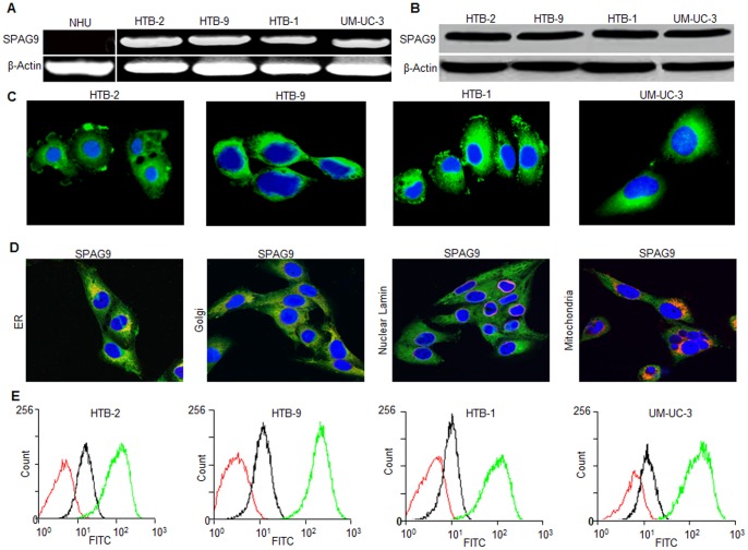
Figure 6. SPAG9 expression in bladder cancer cell lines.
(A) SPAG9 mRNA expression assessed by RT-PCR revealed the presence of SPAG9 transcript in all bladder cancer cells. However, NHU cells did not reveal SPAG9 transcript. β-Actin was used as an internal control. (B) Western blot analysis of SPAG9 protein expression. (C) Indirect immunofluorescence revealed SPAG9 cytoplasmic localization in all bladder cancer cells. Nuclear staining was done by DAPI. (D) UM-UC-3 cancer cells were probed for SPAG9 and various markers for cell organelles. Co-localization of SPAG9 was examined by indirect immunofluorescence assay which revealed SPAG9 co-localization with ER and Golgi marker. Nuclear envelop and mitochondria did not show co-localization with SPAG9. Immunofluorescence staining was detected by a laser-scanning confocal microscope. The images are merged for co-localization of SPAG9 (green) and marker co-staining (red). Original magnification, ×630; objective, 63×). (E) Flow cytometric analysis demonstrated surface localization of SPAG9 protein in fixed cancer cells (green histogram) compared with cells probed with control IgG (black histogram) or stained with secondary antibody only (red histogram). Results from 1 of 3 representative experiments are shown.

