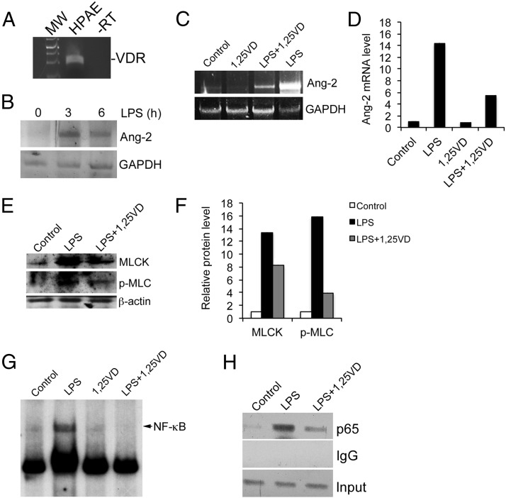
Figure 5.
1,25(OH)2D3 down-regulates Ang-2 by targeting NF-κB activation in HPAE cells. A, RT-PCR detection of human VDR mRNA transcript in HPAE cells. −RT, minus reverse transcriptase control. B, HPAE cells were treated by LPS for 3 and 6 hours, and Ang-2 mRNA induction was measured by RT-PCR. C and D, HPAE cells were treated with LPS in the presence or absence of 1,25(OH)2D3 (20 nM). Ang-2 mRNA was assessed by RT-PCR at 24 hours (C) and semiquantified by densitometry (D). E and F, HPAE cells were treated with LPS in the presence or absence of 1,25(OH)2D3. MLCK and phosphorylated MLC levels were assessed by Western blotting after 24 hours (E) and semiquantified by densitometry (F). G, EMSA. HPAE cells were treated with LPS in the presence or absence of 1,25(OH)2D3 for 24 hours. Nuclear extracts were prepared for EMSA using 32P-labeled putative κB probe within the first intron of human ANG-2 gene. H, ChIP assay. HPAE cells were treated with LPS in the presence or absence of 1,25(OH)2D3 for 6 hours. ChIP assays were performed using anti-p65 antibodies or nonimmune IgG. PCR amplification was performed using primers flanking the putative κB site within the first intron of human ANG-2 gene. GAPDH, glyceraldehyde-3-phosphate dehydrogenase; MW, molecular weight; −RT, minus reverse transcriptase; 1,25VD, 1,25(OH)2D3; p-MLC, phosphorylated MLC.

