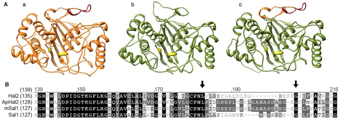
Figure 4. Modelling of a modified SAL1 protein with the inserted loop with the META region from ApHal2.

Top: Three-dimensional models of A. pullulans ApHal2 (A), A. thaliana SAL1 (B), and modified SAL1 (mSAL1) (C) with the replaced loop shown in orange and the META region in red. The conserved active-site amino acids are in yellow. The models were prepared on the basis of the S. cerevisiae Hal2 structure, using Swissmodel. Bottom: Amino-acid alignment of Hal2 (NP_014577), ApHal2 (KC242234), mSAL1 and SAL1 (ID 836519), using AlignX. Black arrows indicate the conserved amino acids lysine (170) and glycine (189), bordering on the loop of the SAL1 protein, which was replaced by the loop from ApHal2, containing the META region (DSEPLTEDL).
