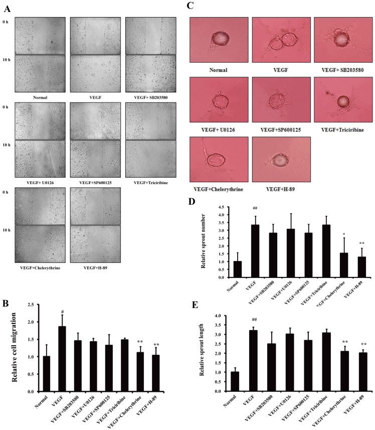
Figure 3. The role of the PKA pathway in the VEGF-induced endothelial cell migration.
(A). Effects of specific inhibitors of various pathways on wound healing. Scratch wounds were created in cell monolayers of HUVECs using a sterile pipette tip. Then cells were cultured with inhibitors (10 µM) in the presence of VEGF (10 ng/ml) for 10 h. (B). Quantification of wound area (The initial wound area minus wound area after 10 h). (C). Effects of specific inhibitors of various pathways on VEGF-induced sprouting of endothelial-cell-coated beads in fibrin gel. Cells on beads were exposed to different treatments, and photographs were taken on day 14. (D). Quantification of sprout number. (E). Quantification of sprout length. The data are expressed as mean ± SD of three independent experiments. # P<0.05, ## P<0.01 vs. normal group; *P<0.05, **P<0.01 vs. VEGF group. (original magnification: ×100 in A, ×200 in B).

