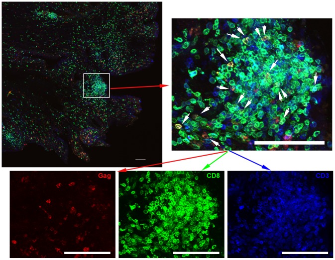
Figure 4. Most CD3+ T cells were CD8+ in genital lymphoid aggregates.
Representative images of endocervix section stained with Mamu-A1/Gag tetramers (red), CD8 antibodies (green), and CD3 antibodies (blue) from animal #338.02. Tetramer+ cells are indicated with white arrows in the enlargement. The bottom panels show individual tetramer and antibody stains. Note most CD3+ T cells are CD8+. The top left panel shows a montage of projected z-series of several 200× fields. Scale bars = 100 µm.

