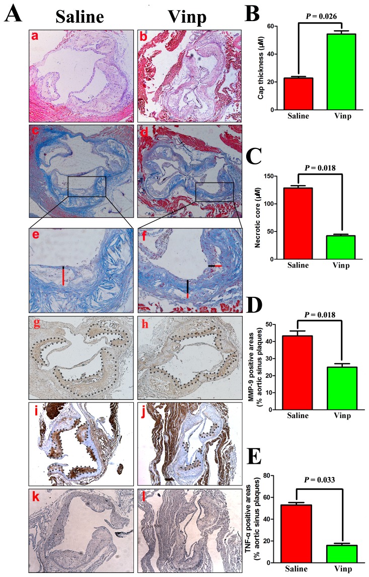
Figure 2. Vinpocetine affects plaque composition in apoE-/- mice.
A. Representive pictures of hematoxylin and eosin staining (a-b), Masson’s trichrome staining (c-f), MMP-9 (g-h), TNF-α (i-j) and normal IgG (k-l) of atherosclerotic plaques in aortic sinus. Through immunohistochemistry, brown staining exhibits positive area, while blue represents counterstaining with hematoxylin. Absolute values of necrotic core size (B) and cap thickness (C) with max depth in local plaques from the aortic sinus and representative images (e and f) to show how the necrotic core (red line) and fibrous cap (black line) in plaques from the aortic sinus was measured. Results represent the percentage of areas occupied by MMP-9 (D) and TNF-α (E) versus total plaque area within aortic sinus. Immunohistochemical staining of normal IgG (k-l) was presented as negative control experiments.
Data are obtained from six mice from each group and bars indicate mean ± SEM.

