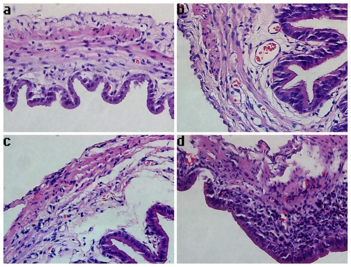
Figure 2. Photomicrographs of gallbladder samples stained with hematoxylin and eosin in each group.
The magnification of photomicrographs is ×400. (a) Cholecystic pathology of a representative sample from the group that underwent the sham operation, showing normal gallbladder histology. (b) Cholecystic pathology of a representative sample of an animal from the CBDL-1 group, indicating edema, congestion, and a few inflammatory cells infiltrating into the mucosa. (c) Cholecystic pathology of a representative sample of an animal from the CBDL-2 group, indicating pathological changes of gallbladder resembling that of the CBDL-1 group. (d) Cholecystic pathology of a representative sample of an animal from the CBDL-3 group, exhibiting pronounced edema with significant inflammatory cell infiltrates in the lamina propria.

