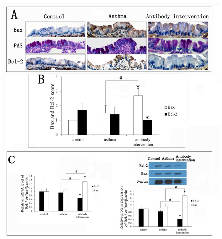
Figure 5. Effects of mCLCA3 antibody on apoptosis-associated genes.
(A) Bcl-2 and Bax expression in goblet cells was measured by immunohistochemistry (magnification x400). (B) Quantitative analyses of Bcl-2 and Bax expression levels in goblet cells. (C) The levels of Bcl-2 and Bax mRNA were detected by RT-PCR. (D) The expression of Bcl-2 and Bax in all groups of mice was analyzed by Western blotting. Bcl-2 and Bax expression levels were normalized with β-actin values and are expressed as fold changes compared to control. Data are shown as mean± SEM, n = 5 (*P<0.05, compared with the control group; #P<0.05, compared with the asthma group).

