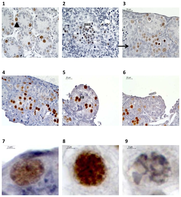
Figure 7. Histological evaluation of meiotic process evaluation after Tra98 immunostaining of organotypic culture of fresh and frozen-thawed pre-pubertal mice testicular tissue.
(1-2-3) Meiotic germ cells assessment for organotypic culture of fresh pre-pubertal mice testicular tissue at ×500 magnification. Fresh testicular tissue was culture for 0 (1) or 11 days (2 and 3) with RE6 (2) or RERA5 (3)
(4-5-6) Meiotic germ cells assessment for organotypic culture of frozen-thawed (5 and 6) immature mice testicular tissue compared to fresh control (4) at ×500 magnification. Testicular tissue was frozen using a temperature stabilisation phase at –8°C (5) or –9°C (6)
(7-8-9) Germ cells classification after Tra98 immunodtection at x1000 magnification.
Intra-tubular cells were classified as spermatogonia (7, arrow head) (smooth spherical brown nuclei), leptotene/zygotene primary spermatocytes (8, ###) (irregular spherical brown nuclei with condensed chromatin) or pachytene primary spermatocytes (9, ¤¤¤) (irregular spherical brown nuclei with highly condensed chromatin). Sertoli cells was defined with a blue nucleus (head).
Footnotes:
RE6: 10-6M retinol ; RERA5: 10-5M retinoic acid and 3.3.10-7M retinol

