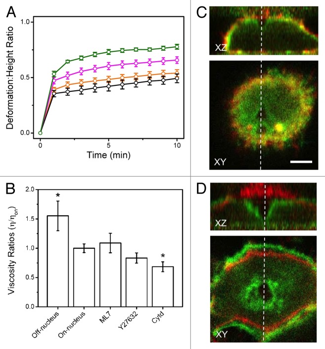
Figure 2. Resistance to deformation is dependent on the cytoskeleton. (A) Deformation: height ratios over time comparing untreated HeLa (orange) to cells treated with ML7 (black), Y27632 (magenta), and Cytd (green). Cells treated with Y27632 and Cytd deformed significantly more than untreated cells. (B) Viscosity ratios derived from Kelvin-Voigt fits of time-dependent strain. Shown are the viscosities of treated cells relative to untreated cells (above the nucleus), as well as the cytoplasmic regions relative to the nuclear region (both untreated). The cytoplasmic regions are more viscous than the nuclear regions, suggesting that they are densely packed with cytoskeletal filaments. As well, cells treated with Cytd are significantly less viscous than untreated cells, indicating that the actin cytoskeleton is mainly responsible for resistance to deformation. Error bars are standard error. (*) indicates p < 0.05 significance compared with on-nucleus results as determined by a t-test. (C) Image overlay of untreated HeLa cells during (after 10 min of 10nN) (green) and prior-to deformation (red). (D) Image overlay of Cytd-treated HeLa cell during (green) and prior-to deformation (red). The deformation is much more pronounced in comparison to untreated cells. Figure adapted from reference 16.

An official website of the United States government
Here's how you know
Official websites use .gov
A
.gov website belongs to an official
government organization in the United States.
Secure .gov websites use HTTPS
A lock (
) or https:// means you've safely
connected to the .gov website. Share sensitive
information only on official, secure websites.
