
Figure 3.
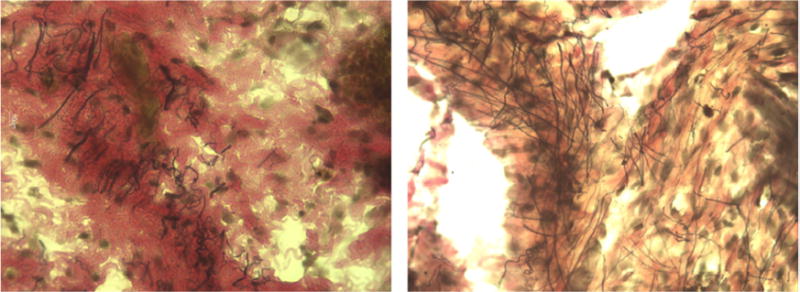
Representative histological section (at 40X) from the upper vaginal wall (left) two days and (right) two weeks postpartum of spontaneous vaginal delivery Sprague-Dawley rats. Elastic fibers are stained black, collagen is stained red, and smooth muscle is stained yellow.