
Figure 4.
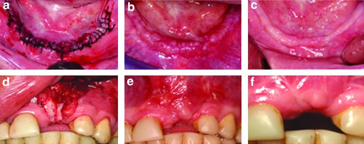
(a) Lower jaw of a 62-year-old male patient at the day of oral surgery. Silk sutures (black) were placed to close the large incisional wound created to the oral and gingival tissues for the removal of four teeth, insertion of two dental implants, and placement of bone grafts. The same surgical area is shown at (b) 2 weeks and (c) 4 months postoperatively. No scar tissue was clinically observed. (d) Insertion of a free keratinoconjuntival graft tissue (to augment the lack of keratinized tissue at future implant site). The same surgical area is shown at (e) 2 weeks and (f) 3 months postoperatively. The amount of postoperative scarring is low, considering the extent of the surgical procedure compared to a similar type of skin-grafting surgery. To see this illustration in color, the reader is referred to the web version of this article at www.liebertpub.com/wound